Could Laboratory Developed Lung Tissue Help Asthmatics?
 The development of some of the most realistic lung tissue ever could change the way medication for asthma and chronic obstructive pulmonary disorder (COPD) is researched.
Currently, the safety and effectiveness of drugs for humans are mostly tested using chemical tests or animals or on 2D cell cultures that don’t closely replicate real human organs.
Chemical testing can miss things that may affect a human organ or cells and testing on animals is an unpopular method and often causes outrage, particularly among animal rights activists.
But scientists from Rice University in the United States, and its spinoff company Nano3D Biosciences, have created tissues that are virtually identical to those found in people’s bodies, meaning testing on animals and using chemical testing could be a thing of the past.
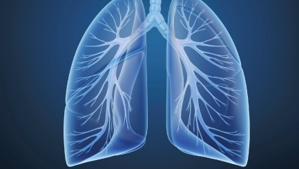
The team of researchers combined four types of cells to replicate tissue from the wall of the bronchiole in the lung, the part of the breathing system that becomes dangerously inflamed and damaged in asthma sufferers.
Historically, laboratory tests have been conducted on flat, 2D cell cultures grown in a flat Petri dish which may not give the same results as a human organ.
The scientists from the United States used magnets to develop a more lifelike, 3D tissue that acts more like a human organ.
Being able to test on cells that replicate a human lung so closely can have huge benefits in toxicological and pharmaceutical testing and help reduce the costs of these tests.
While testing medication on animals may be considered wrong, the development of safe and effective treatment for people with medical conditions is vital. But with the development of more realistic human cells in laboratories, the days of using of animals for testing could be numbered.


Comments are closed.