

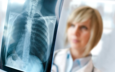
X-rays don’t hurt and are a quick way of diagnosing and monitoring various health conditions. These procedures are carried out by radiographers, who are health professionals trained to carry out these imaging techniques. Generally, an x-ray is performed so that medical professionals can look at the skeletal structure, such as fractures or breaks in bones after an injury. However, many people don’t realise that they can also be used to look at the health of your organs as well. For example, an x-ray of your chest can show whether there is any infection in your lungs. They work as a form of radiation, however unlike light radiation which is absorbed or reflected by your skin, x-ray radiation passes through the body – the x-ray machine works by projecting a beam of rays through the part of your body being examined and this is then captured in the form of a black and white image. This image is known as a radiograph. The structures inside the body which are dense absorb the x-rays and show up as white – for example, your bone structure. Less dense materials, such as air in the lungs, will pass through almost completely and show up as black on the image. The various aspects of your body alter in density, so the radiograph will show various parts in shades of grey.
It depends which area of your body is being looked at, but a different type of imaging procedure may be recommended as more appropriate than an x-ray. For example, you may need an ultrasound scan, a CT scan or an MRI scan. Your doctor will advise which is needed. The x-ray procedure is generally done as an out-patient procedure in the radiology department at hospitals. Your radiographer will discuss the process with you beforehand, so that you know what to expect. If you have any questions about what will happen during the procedure, this is an ideal time to ask so that your radiographer can talk you through any concerns you have. They don’t usually take longer than a few minutes to carry out, and depending on the area being examined, you may be asked to remove your clothing and jewellery, and wear a hospital gown instead. Once the procedure has been carried out, a report will be sent to your doctor outlining any problems – this can take a few days.
There are, of course, some risks attached to an x-ray, as with any procedure. The benefits of this imaging technique usually outweigh the risks by a long way, though. Due to the nature of the process, you will be exposed to some x-ray radiation, but the amount isn’t considered harmful and you will only be exposed to it for a few minutes. However, this varies depending on the type of x-ray you’re having – for example, a chest x-ray will expose you to a very small amount of radiation, which is no more than you would be exposed to naturally over the course of about two to three days. But there are other tests for various ailments which leave you exposed to more, such as a barium enema or a barium swallow and meal, which involve having many images taken. Your radiographer is trained to limit the amount of radiation you’re exposed to to a minimum though, so there should be little cause for concern. This situation varies slightly for pregnant women though, as the risk to unborn babies is slightly higher. Unless there is an urgent medical reason, x-rays are rarely carried out on pregnant women – there will be a lead shield to protect your abdomen to help protect your baby if you do need to have an x-ray. If you think you might be pregnant, it’s important to tell your doctor before your appointment – they will then advise if it is safe for you to go ahead with the procedure.
Comments are closed.